Orbital Atherectomy: Procedure, Benefits, Risks and Recovery
Quick Answer
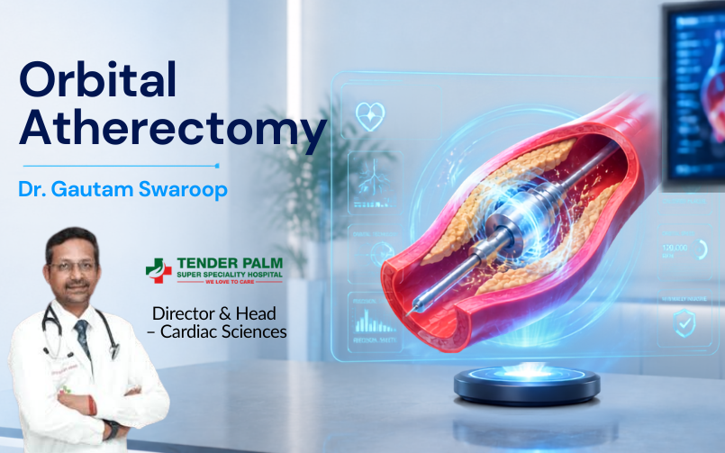
What is orbital atherectomy? Orbital atherectomy is a minimally invasive interventional cardiology procedure used to treat severely calcified coronary arteries. The procedure utilizes a specialized catheter with a spinning, diamond-coated crown to safely sand away hard calcium buildup into microscopic particles. Consequently, this plaque modification allows for the successful expansion and deployment of a coronary stent to restore normal blood flow.
Introduction
When managing advanced coronary artery disease, standard treatments like balloon angioplasty can run into significant roadblocks. Specifically, when plaque inside the heart vessels hardens into dense calcium deposits, standard balloons often cannot expand properly. If your cardiologist recently mentioned that your heart blockages are heavily calcified, you are likely looking into advanced plaque modification options such as orbital atherectomy.
At Dr Gautam Swaroop’s private clinic, we prioritize patient education alongside cutting-edge cardiovascular interventions. Severely calcified blockages act like concrete walls inside your blood vessels, making routine stent placement highly challenging. Fortunately, modern interventional cardiology offers precise, mechanical tools designed to modify this stubborn tissue safely from the inside out.
In this comprehensive guide, we will break down exactly how orbital atherectomy works, why calcified arteries demand specialized care, the step-by-step procedure, and how this technique compares to alternative technologies like rotational atherectomy and shockwave therapy (IVL). Ultimately, this guide will help you understand your options for achieving optimal long-term heart health.
Table of Contents
- What Is Orbital Atherectomy?
- Why Do Calcified Coronary Arteries Require Special Treatment?
- How Does Orbital Atherectomy Work?
- Who Needs Orbital Atherectomy?
- Orbital Atherectomy Procedure Step by Step
- Benefits of Orbital Atherectomy
- Risks and Complications
- Orbital Atherectomy vs Rotational Atherectomy
- Orbital Atherectomy vs Intravascular Lithotripsy (IVL)
- Recovery After Orbital Atherectomy
- Success Rates and Long-Term Outcomes
- When Should You Consult an Interventional Cardiologist?
- Key Takeaways
- Frequently Asked Questions
- Conclusion
What Is Orbital Atherectomy?
Definition Box: Orbital atherectomy is an advanced, catheter-based percutaneous coronary intervention (PCI) that utilizes a high-speed, eccentrically mounted, rotating diamond-coated crown to mechanically shave away calcified plaque from the interior walls of coronary arteries.
Unlike traditional angioplasty which attempts to push plaque aside using fluid pressure, this specialized system alters the mechanical properties of the vessel wall. By sanding down the calcified structure, the procedure softens the artery. As a result, the vessel becomes flexible enough to receive a permanent coronary stent safely.
Why Do Calcified Coronary Arteries Require Special Treatment?
Coronary artery disease typically begins as a soft accumulation of cholesterol and fat. However, as the disease progresses over several years or decades, the body deposits calcium into this fibrous matrix, essentially turning the soft plaque into a bone-like structure. When severe calcification occurs, it poses unique challenges during a standard angioplasty procedure.
Challenges of Severe Artery Calcification
The presence of hard calcium inside a coronary artery creates multiple clinical hurdles. Understanding these challenges explains why interventional cardiologists rely on specialized tools:
| Clinical Challenge | The Risk Factor | The Advanced Plaque Solution |
|---|---|---|
| Inadequate Balloon Expansion | Standard angioplasty balloons rupture or fail to expand symmetrically against rigid, calcified walls. | Mechanical sanding breaks the rigid calcium rings. |
| Stent Underexpansion | If a stent cannot fully deploy, it leaves a narrow channel, vastly increasing the risk of sudden blood clots (stent thrombosis). | Modifying the plaque ensures the stent expands to its maximum designed diameter. |
| Vessel Damage | Applying extreme high pressure via standard balloons to crack calcium can cause unpredictable tears or vessel ruptures. | A rotating crown delivers controlled, low-impact mechanical friction strictly to calcified areas. |
How Does Orbital Atherectomy Work?
The mechanics of orbital atherectomy rely on a balance of physics and bio-compatible engineering. The entire procedure operates on a fundamental principle known as differential sanding.
The Orbital Atherectomy System
The system consists of a handheld electric controller and a specialized catheter sheath. This sheath houses an ultra-thin guide wire over which a drive shaft operates. At the working tip of this shaft sits an eccentric (off-center) diamond-coated crown.
Diamond-Coated Crown Technology
Because the diamond-coated crown is mounted off-center, it does not just spin in place like a drill bit. Instead, as the rotation speed increases, centrifugal force drives the crown outward. Consequently, the crown moves in an expanding orbital path, similar to a lasso swinging in a circle or a planet orbiting the sun. This orbital movement ensures that the device can treat a vessel diameter much larger than the catheter tip itself.
Plaque Modification Process
The unique mechanism safely differentiates between healthy and diseased tissue. Here is the step-by-step mechanical process flow:
- Tissue Selection: When the spinning crown touches soft, elastic healthy arterial tissue, the healthy tissue simply deflects away harmlessly.
- Sanding Hard Calcium: When the crown encounters rigid, inelastic calcified plaque, it cannot deflect. Therefore, the micro-diamonds grind away the surface layer of the calcium.
- Particle Elimination: The friction grinds the hard plaque into microscopic debris particles. These fragments measure less than 2 microns in diameter, which is significantly smaller than a red blood cell.
- Safe Clearance: Because the particles are incredibly small, they pass harmlessly through the heart’s microscopic capillary network and get cleared naturally by the body’s reticuloendothelial system, preventing blockages in downstream microvessels.
Who Needs Orbital Atherectomy?
Cardiologists do not use orbital atherectomy for routine, soft fatty blockages. Instead, they reserve this advanced intervention for complex patient profiles exhibiting severe calcification patterns confirmed via advanced coronary imaging tools.
Patient Eligibility Profile
| Highly Eligible Candidates | Ineligible Candidates (Contraindications) |
|---|---|
| Patients with severe coronary calcification confirmed via Intravascular Ultrasound (IVUS) or Optical Coherence Tomography (OCT). | Patients with highly tortuous (severely twisted or bent) blood vessels where the rigid device wire cannot pass safely. |
| Individuals experiencing recurrent angina (chest pain) despite optimized medical therapy. | Patients with an active dissection (an existing tear) in the target coronary artery wall. |
| Patients with complex, long segment blockages where standard balloons cannot expand or pass. | Patients with severe multi-vessel disease who are better suited for open-heart Coronary Artery Bypass Grafting (CABG). |
Orbital Atherectomy Procedure Step by Step
The procedure takes place in a cardiac catheterization laboratory (cath lab). Dr Gautam Swaroop performs this intervention with precision, utilizing continuous real-time imaging.
The Clinical Timeline
- Sedation and Local Anesthesia: The clinical team administers mild intravenous sedation to keep you relaxed and comfortable. They apply local anesthesia to numb the access site, typically in the wrist (radial artery) or the groin (femoral artery).
- Catheter Introduction: The interventional cardiologist inserts a hollow guiding catheter into the artery and carefully advances it up to the openings of the heart’s coronary arteries under fluoroscopic (X-ray) guidance.
- Lesion Assessment via Imaging: Before using the ablation device, the doctor uses advanced intravascular imaging (such as IVUS or OCT) to map out the exact depth, thickness, and arc of the calcium deposits.
- Device Positioning: The physician advances a specialized, ultra-thin atherectomy guide wire across the calcified blockage. They then slide the orbital atherectomy catheter over this wire, placing the diamond crown just before the blockage.
- Controlled Plaque Ablation: The doctor activates the device. The crown spins at a baseline speed of 80,000 RPM, gently advancing across the blockage for up to 20 seconds per pass. If needed, the doctor can increase the speed to 120,000 RPM to sweep a wider orbital path. Continuous fluid infusion lubricates and cools the device during activation.
- Coronary Stent Placement: Once the calcium ring is successfully fractured and modified, the doctor removes the device. They insert a standard angioplasty balloon to open the channel easily, followed by the deployment of a permanent drug-eluting coronary stent.
- Final Verification and Closure: The physician performs a final intravascular imaging sweep to verify that the stent has expanded perfectly against the vessel wall. They remove all catheters and apply a closure device or compression band to the entry site to ensure proper healing.
Benefits of Orbital Atherectomy
Utilizing orbital atherectomy dramatically improves the safety and success rates of percutaneous coronary interventions in challenging, bone-dry calcified lesions.
Key Benefits Summary
| Advantage | Clinical Impact |
|---|---|
| Optimal Stent Apposition | Ensures the metal stent presses completely flat against the artery wall, minimizing the risk of long-term restenosis (re-narrowing). |
| Continuous Blood Flow | The small crown profile allows blood to continue flowing around the device even while it is spinning, lowering the risk of procedural chest pain. |
| Bi-Directional Sanding | The diamond crown sands plaque while moving both forward and backward, drastically reducing the risk of the device getting stuck inside a tight blockage. |
| Fewer Vascular Complications | Controlled mechanical sanding prevents the need for high-pressure balloon inflation, reducing the incidence of severe arterial tears. |
Risks and Complications
While orbital atherectomy is highly refined, performing any mechanical ablation inside a delicate heart artery carries certain medical risks. Interventional cardiologists weigh these risks carefully against the necessity of clearing the vessel.
- Coronary Artery Perforation: A rare but severe risk where the spinning crown cuts completely through the blood vessel wall, requiring immediate repair via a covered stent or emergency surgery.
- Slow-Flow or No-Reflow Phenomenon: Microscopic debris particles can temporarily clog downstream capillaries, causing a brief drop in blood flow and potential rhythm disturbances.
- Thermal Injury: High-speed rotation generates friction heat. However, the system minimizes this risk by mandating constant infusion of a specialized cooling cocktail solution during activation.
- Abrupt Vessel Closure: The treated artery could potentially spasm or collapse immediately after ablation, requiring swift balloon inflation or stent rescue.
- Access Site Complications: Minor bleeding, bruising, or hematoma formation can occur where the catheter entered the radial or femoral artery.
Orbital Atherectomy vs Rotational Atherectomy
Interventional cardiologists frequently choose between orbital atherectomy and rotational atherectomy depending on the specific anatomy of the blockage.
| Feature | Orbital Atherectomy (OA) | Rotational Atherectomy (RA) |
|---|---|---|
| Crown/Burr Design | Eccentric (off-center) diamond-coated crown. | Concentric (centered) football-shaped diamond burr. |
| Ablation Mechanism | Orbital path; orbits wider at higher speeds. Trepanning action. | Drills a fixed-diameter channel straight through the center. |
| Sanding Movement | Bi-directional (cuts moving forward and backward). | Uni-directional (cuts only moving forward; risk of trapping). |
| Vessel Size Flexibility | A single catheter size can treat varying vessel diameters by adjusting speed. | Requires upsizing to larger burrs manually for wider vessels. |
| Ischemic Protection | Higher; blood flows around the crown during activation. | Lower; the burr can completely occlude the vessel while drilling. |
Orbital Atherectomy vs Intravascular Lithotripsy (IVL)
Another major alternative for plaque modification is Intravascular Lithotripsy (IVL), commonly referred to as shockwave therapy.
| Feature | Orbital Atherectomy (OA) | Intravascular Lithotripsy (IVL) |
|---|---|---|
| Primary Modality | Mechanical ablation (sanding away surface tissue). | Acoustic shockwaves (cracking deep calcium layers). |
| Target Location | Most effective for superficial, concentric calcium lining the inner lumen. | Highly effective for deep, thick, eccentric, or medial calcium. |
| Device Profile | Thin wire and crown profile; can cross ultra-tight, pinhole narrowings. | Bulkier balloon profile; requires a wider opening just to pass the lesion. |
| Tissue Debulking | Physically removes tissue volume from inside the vessel. | Does not remove tissue; fractures the ring structure to allow stretching. |
Recovery After Orbital Atherectomy
The post-operative recovery timeline is surprisingly swift, matching standard angioplasty protocols. After the catheters are removed, you will spend several hours in a specialized recovery area.
If your procedure used a radial entry site in your wrist, you can typically sit up and eat shortly after completion. Most patients remain in the hospital overnight for observation and return home the following morning. You must strictly avoid heavy lifting, vigorous exercise, or driving for approximately 5 to 7 days to give the arterial puncture site time to heal completely.
Furthermore, following stent placement, you must strictly comply with your prescribed Dual Antiplatelet Therapy (DAPT)—usually a combination of aspirin and a medication like clopidogrel or ticagrelor. Stopping these medications without explicit permission from Dr Gautam Swaroop can cause life-threatening blood clots to form inside the new stent.
Success Rates and Long-Term Outcomes
Extensive clinical trial data confirms that orbital atherectomy delivers outstanding success rates when managing complex, calcified heart disease. By thoroughly preparing the vessel wall, the procedure ensures that over 90% of cases achieve optimal stent expansion. Long-term studies show that modifying plaque with this system significantly minimizes target lesion revascularization (the need for a repeat procedure) and reduces the long-term risk of major adverse cardiac events (MACE). Ultimately, it provides a durable, reliable solution for complex blockages.
When Should You Consult an Interventional Cardiologist?
If you have known coronary artery disease, you should monitor your symptoms closely. You should arrange a formal evaluation at Dr Gautam Swaroop’s clinic if you experience:
- Persistent chest pain, pressure, or tightness (angina) that occurs during minimal physical activity.
- Shortness of breath that limits your daily functional walking capacity.
- Dizziness, unexplained lightheadedness, or brief fainting spells.
- A history of coronary blockages where you were previously told that standard stenting was “too high-risk” or impossible due to hard plaque.
Key Takeaways
- Orbital atherectomy is an advanced interventional cardiology procedure used to modify severely calcified coronary arteries.
- The system utilizes an off-center, diamond-coated crown spinning at high speeds (up to 120,000 RPM).
- It relies on centrifugal force to create an expanding orbital path, allowing a single catheter to treat various vessel sizes.
- The procedure sands away hard calcium into microscopic particles smaller than a red blood cell.
- Modifying rigid calcium ensures that a coronary stent can expand fully, preventing long-term stent failure.
- Unlike rotational devices, this system offers bi-directional cutting and allows continuous blood flow during activation.
- Recovery typically requires a single overnight hospital stay and a brief week-long restriction on heavy physical lifting.
- Strict adherence to post-procedure blood thinners (DAPT) is mandatory to ensure long-term safety.
Frequently Asked Questions
1. What is the main purpose of orbital atherectomy?
The primary purpose is plaque modification. It mechanically sands down hard calcium deposits lining the walls of coronary arteries, creating cracks in the rigid structure so a coronary stent can deploy successfully.
2. Is orbital atherectomy considered open-heart surgery?
No. It is a minimally invasive percutaneous coronary intervention (PCI) performed through a tiny puncture in the wrist or groin inside a cardiac catheterization laboratory.
3. How does the device avoid damaging healthy parts of the artery?
The system relies on differential sanding. Elastic, healthy arterial tissue simply flexes away from the spinning diamond crown without injury, whereas hard, rigid calcium cannot move and gets sanded down.
4. What happens to the sanded plaque particles?
The diamond crown pulverizes the calcium into microscopic fragments smaller than 2 microns. These fragments pass harmlessly through the heart’s capillary bed and are cleared naturally by the body.
5. What is the difference between orbital and rotational atherectomy?
Orbital atherectomy uses an off-center crown that cuts in both directions and allows blood flow during use. Rotational atherectomy uses a centered, football-shaped burr that drills straight forward like a drill bit.
6. Does the procedure require general anesthesia?
No. Doctors use local anesthesia to numb the entry site and mild intravenous sedation to help you stay relaxed. You remain awake and communicative throughout the process.
7. How long does the entire procedure take?
The atherectomy portion adds only a few minutes to a procedure. However, the entire cardiac catheterization, imaging, and stenting process generally takes between 1 to 2 hours.
8. What is the normal recovery time after going home?
Most patients return to light daily routines within 48 hours. However, you must avoid strenuous exercise or heavy lifting for about 5 to 7 days to protect the entry wound site.
9. Can the device get permanently stuck inside the heart artery?
The risk of device entrapment is extremely low because the eccentric crown sands moving both forward and backward, allowing the cardiologist to retract it safely if it encounters resistance.
10. What are the most common risks of this procedure?
Potential risks include vascular perforation, brief drops in blood flow (slow-reflow), arterial spasms, or standard catheterization risks like minor bleeding at the entry site.
11. Who is an ideal candidate for an orbital atherectomy test?
Ideal candidates are patients with advanced coronary artery disease showing severe concentric calcification on intravascular imaging (IVUS/OCT) who require stent placement.
12. Can a patient undergo this procedure if they have severe asthma?
Yes. Unlike rotational techniques which sometimes rely on specific medications that affect the airways, orbital atherectomy does not carry specific respiratory contraindications.
13. How durable are the long-term results of this procedure?
Long-term outcomes are excellent. By ensuring full stent expansion, the procedure significantly reduces the likelihood of the stent narrowing again or failing in the future.
14. Do I need to take special medications after the procedure?
Yes. You must take Dual Antiplatelet Therapy (DAPT), consisting of aspirin and a secondary blood thinner, exactly as prescribed to keep your new stent clear and open.
15. Is orbital atherectomy covered by health insurance medical policies?
Yes. Most major corporate and private health insurance providers cover advanced plaque modification procedures when intravascular imaging documents severe calcification.
Conclusion
Navigating advanced coronary artery disease demands highly specialized solutions. By understanding how orbital atherectomy acts as an advanced plaque modification tool, you can see how modern interventional cardiology safely overcomes the challenges of severe calcification. Grinding down rigid calcium barriers restores elasticity to your blood vessels. Ultimately, combining precise mechanical technology with coronary stent placement allows patients to break free from chronic chest pain and regain an excellent, highly active quality of life.
Book your Appointment now
Are you or a loved one dealing with severe coronary artery disease, complex blockages, or recurrent angina? Don’t let calcified arteries stand in the way of your recovery. Contact Dr Gautam Swaroop today to schedule a comprehensive evaluation. As an expert interventional cardiologist, Dr Gautam Swaroop specializes in advanced plaque modification techniques like orbital atherectomy, providing personalized, cutting-edge solutions to restore your heart’s health safely. Visit drgautamswaroop.in to book your consultation.

You must be logged in to post a comment.