Catheter-Based Repair for Congenital Heart Defects: A Complete Guide to Minimally Invasive Treatment
Quick Answer
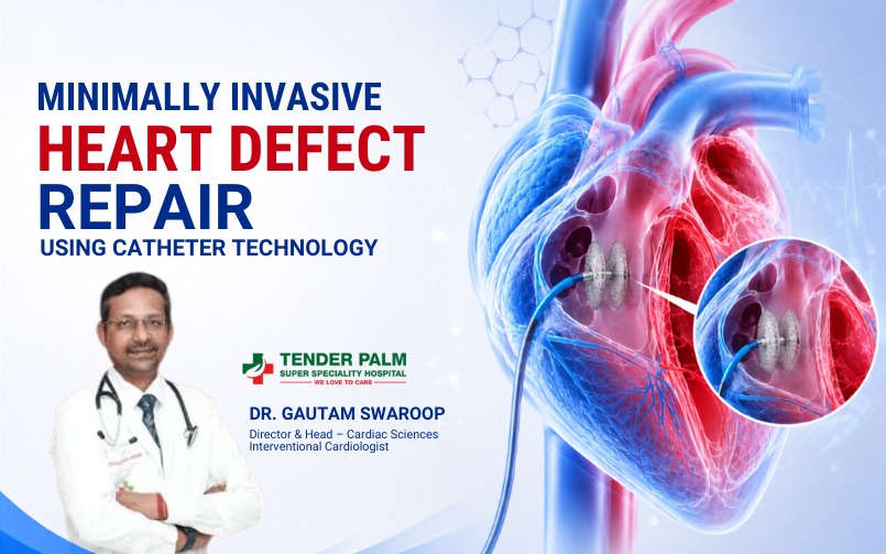
Catheter-based repair for congenital heart defects is a minimally invasive way to treat selected heart defects without opening the chest. Instead of traditional open-heart surgery, an interventional cardiologist passes a thin, flexible tube called a catheter through a blood vessel, usually in the groin, and guides it to the heart using live imaging. Through that catheter, the doctor places a device, plug, balloon, or stent to close a hole or widen a narrowed valve or blood vessel.
This treatment is commonly used for selected defects such as atrial septal defect (ASD), patent ductus arteriosus (PDA), pulmonary valve stenosis, some ventricular septal defects (VSDs), and selected cases of coarctation of the aorta. It is often preferred when the heart anatomy is suitable and the patient is a good candidate for a non-surgical repair.
The main benefit is simple: less trauma, less pain, smaller scars, shorter hospital stay, and faster recovery. But it is not suitable for every defect, and specialist evaluation is always necessary before deciding the right treatment.
Introduction
Congenital heart defects are structural problems of the heart that are present from birth. Some are small and never cause symptoms. Others can affect blood flow, strain the heart, reduce oxygen delivery, and lead to long-term complications if they are not treated in time. In children, these defects may show up as feeding difficulty, poor weight gain, fast breathing, or repeated chest infections. In adults, they may appear as tiredness, breathlessness, palpitations, or a heart murmur found during a routine exam.
The good news is that modern cardiology has changed the way many of these problems are treated. For selected congenital heart defects, treatment no longer means a large chest incision or a long surgical recovery. Catheter-based repair now allows doctors to treat many structural heart problems through a small puncture in the skin. This is one of the most important advances in modern heart care because it can reduce pain, shorten recovery, and help patients return to normal life sooner.
At the same time, this treatment is not a shortcut for every case. Some heart defects are simple and suitable for catheter closure. Others are too complex, too large, or too close to important valves or vessels, and still need open-heart surgery. That is why careful diagnosis is so important. The right imaging, the right specialist, and the right timing make all the difference.
This guide explains what congenital heart defects are, how catheter-based repair works, which defects can be treated this way, who is a good candidate, what to expect during the procedure, and how recovery usually goes. It also compares catheter repair with open-heart surgery in a practical and honest way.
Table of Contents
- What Are Congenital Heart Defects?
- How Are Congenital Heart Defects Diagnosed?
- What Is Catheter-Based Repair?
- Which Congenital Heart Defects Can Be Treated Using Catheter-Based Procedures?
- Who Is a Good Candidate?
- Preparing for a Catheter-Based Procedure
- Step-by-Step Procedure
- Benefits of Catheter-Based Repair
- Risks and Possible Complications
- Recovery After Catheter-Based Repair
- Life After the Procedure
- Catheter-Based Repair vs Open-Heart Surgery
- Common Myths About Catheter-Based Repair
- When Should You Consult a Cardiologist?
- Frequently Asked Questions
- Key Takeaways
What Are Congenital Heart Defects?
A congenital heart defect is a problem in the structure of the heart or the large blood vessels connected to it that develops before birth. These defects happen while the baby’s heart is forming in the womb, usually very early in pregnancy. In some cases, the cause is genetic. In others, there may be an association with maternal illness, certain medications, infections, or environmental factors. Often, no exact cause is found.
Congenital heart defects can affect the walls, valves, chambers, or major arteries of the heart. Some are simple, such as a small hole between chambers. Others are complex and involve several abnormalities at once. The severity depends on the type of defect, its size, where it is located, and how much it changes the flow of blood.
Common congenital heart defects include:
- Atrial Septal Defect (ASD): A hole in the wall between the two upper chambers of the heart.
- Ventricular Septal Defect (VSD): A hole in the wall between the two lower chambers.
- Patent Ductus Arteriosus (PDA): A fetal blood vessel that fails to close after birth.
- Pulmonary Valve Stenosis: A narrowing of the valve that sends blood from the right side of the heart to the lungs.
- Coarctation of the Aorta: A narrowing in the main artery that carries blood from the heart to the rest of the body.
Some congenital defects are detected in infancy, but others are found later in childhood or even adulthood. A person may live with a small defect for years without knowing it. That is why a heart murmur, unexplained tiredness, exercise intolerance, or repeated breathing problems should never be ignored.
If a defect is left untreated and is hemodynamically significant, it can lead to enlargement of the heart, pulmonary hypertension, rhythm problems, stroke risk, or heart failure over time. Early diagnosis gives doctors the best chance to intervene before these complications become serious.
How Are Congenital Heart Defects Diagnosed?
Diagnosis usually starts with a doctor’s examination and then moves to imaging.
A heart murmur, breathlessness, poor weight gain, repeated chest infections, fatigue, or low exercise tolerance may make a doctor suspect a congenital problem. The most common tests include:
- Physical examination: The doctor listens for a murmur, checks oxygen levels, and looks for signs of heart strain.
- Echocardiography: This is the most important test in many cases. It uses ultrasound to show the heart chambers, valves, blood flow, and defect size.
- Electrocardiogram (ECG): This checks the heart rhythm and may show chamber enlargement or strain.
- Chest X-ray: This can show heart size, lung blood flow, and general chest structure.
- CT scan or MRI: These are used when anatomy is more complex or more detail is needed before planning catheter repair.
- Cardiac catheterization: In selected cases, this gives exact pressure measurements and helps decide whether a defect can be safely treated.
This step matters because not all defects that look similar are actually treated the same way. A small change in anatomy can decide whether catheter repair is safe or whether surgery is the better choice.
What Is Catheter-Based Repair?
Catheter-based repair is a minimally invasive way to treat selected congenital heart defects. Instead of opening the chest and operating directly on the heart, the doctor inserts a thin, flexible tube called a catheter into a blood vessel, most often in the groin. This catheter is guided through the bloodstream into the heart or nearby vessels.
The procedure is performed in a cardiac catheterization laboratory using live imaging such as fluoroscopy and echocardiography. These imaging tools help the doctor see the defect clearly and place the repair device in the correct position.
Depending on the defect, the doctor may use:
- A closure device to seal a hole such as ASD or PDA
- A balloon to widen a narrowed valve or blood vessel
- A stent to support a narrowed artery and keep it open
The major advantage of catheter-based repair is that it avoids a large surgical incision and, in many cases, avoids the need for a heart-lung machine. That means the body experiences less trauma. For the right patient, this can mean shorter hospitalization, lower pain levels, quicker recovery, and a faster return to school, work, and daily life.
But the procedure works only when the heart anatomy is suitable. Some defects cannot be closed safely with a catheter. In those cases, surgery remains the better option. That is why the decision must be made by an experienced heart team, not by guesswork.
Which Congenital Heart Defects Can Be Treated Using Catheter-Based Procedures?
Catheter-based treatment is not used for all congenital heart defects. It is most useful for defects where a device or balloon can safely fix the problem without open surgery.
Atrial Septal Defect (ASD)
An ASD is a hole in the wall between the heart’s upper chambers. This allows oxygen-rich blood to move from the left atrium to the right atrium, which increases blood flow to the lungs and can enlarge the right side of the heart.
Small ASDs may not cause symptoms. Larger ones can cause breathlessness, fatigue, palpitations, reduced exercise tolerance, or frequent chest infections. Over time, untreated ASD may increase the risk of atrial arrhythmias, pulmonary hypertension, or stroke.
Catheter closure is commonly done for secundum ASD when the surrounding tissue is suitable for device placement. During the procedure, a closure device is delivered through the catheter and released across the hole. The device has two discs that sit on both sides of the defect, sealing it in place. For suitable patients, this offers excellent outcomes and avoids open-heart surgery.
Patent Ductus Arteriosus (PDA)
The ductus arteriosus is a normal fetal blood vessel that connects the pulmonary artery to the aorta before birth. After birth, it is supposed to close. If it stays open, blood flows abnormally from the aorta into the pulmonary artery, increasing blood flow to the lungs and stressing the heart.
A PDA may be silent when small. Larger PDAs can cause fast breathing, sweating during feeding in infants, failure to thrive, and eventually heart enlargement or pulmonary hypertension.
Catheter-based closure is a standard treatment for many PDAs. A device is placed through the catheter into the open vessel and left permanently in position. Over time, the body grows tissue around it and seals the defect completely. For many patients, this is a straightforward, effective treatment with a fast recovery.
Pulmonary Valve Stenosis
Pulmonary valve stenosis is a narrowing of the valve that carries blood from the right ventricle to the lungs. Because blood has to push through a tight opening, the right side of the heart works harder than normal.
Mild cases may not cause symptoms at all. Moderate or severe cases can lead to breathlessness, fatigue, dizziness, or chest discomfort. In babies, serious narrowing can cause low oxygen levels and poor feeding.
Balloon valvuloplasty is the standard catheter-based treatment in many suitable cases. A balloon is passed across the narrowed valve and inflated briefly to stretch the opening. This improves blood flow and reduces pressure on the heart. In many patients, this is enough to avoid surgery.
Selected Ventricular Septal Defects (VSD)
A VSD is a hole in the wall between the lower chambers of the heart. It allows blood to pass from the left ventricle to the right ventricle, increasing flow to the lungs and making the heart work harder.
Many small VSDs close on their own and need only observation. Larger VSDs can cause poor feeding, fast breathing, failure to thrive in children, and heart failure if left untreated. Adults with a small residual VSD may have few or no symptoms but still require follow-up.
Catheter closure of VSD is possible only in selected cases. It is usually considered when the defect is in a favorable location, especially muscular VSDs or selected residual defects after surgery. Because the anatomy is more complex than ASD or PDA, this procedure requires a highly experienced team. In many cases, open surgery is still the preferred treatment.
Coarctation of the Aorta
Coarctation of the aorta is a narrowing in the main artery that carries blood from the heart to the body. This makes it harder for blood to reach the lower body and causes the heart to pump against higher pressure.
Symptoms may include high blood pressure, weak leg pulses, headaches, leg fatigue, or poor feeding in infants. Severe narrowing in newborns may cause heart failure early in life.
In selected older children, teens, and adults, catheter treatment with balloon angioplasty and sometimes stent placement can widen the narrowed segment. This is especially useful in localized narrowing or in patients who have restenosis after previous repair. In newborns and in complex long-segment narrowing, surgery is usually needed.
Who Is a Good Candidate?
The decision to treat a congenital heart defect with catheter-based repair depends on more than just the diagnosis. The real question is whether the anatomy is suitable.
A good candidate usually has:
- A defect that can be reached safely through a blood vessel
- A size and location that allow device placement
- No major surrounding structures that would be damaged by the device
- A body size that can safely accommodate the procedure
- A heart condition that does not already require open surgery
A multidisciplinary heart team may include an interventional cardiologist, pediatric or adult congenital cardiologist, cardiac surgeon, anesthesiologist, and imaging specialist. This team reviews whether catheter repair is the best option or whether surgery would be safer and more effective.
Some patients are not good candidates for catheter repair. That includes people with unsuitable anatomy, very large defects, severe pulmonary hypertension, active infection, or defects that are too close to important valves or vessels. In those situations, surgery may be the correct choice.
Preparing for a Catheter-Based Procedure
Preparation is simple, but it matters.
Before the procedure, the doctor reviews imaging, medications, allergies, and blood tests. The patient may be asked to stop eating or drinking for several hours before the intervention. If the patient is taking blood thinners or diabetes medicines, the doctor may adjust them temporarily.
It is also important to arrange practical details. Children may need a parent present. Adults may need someone to drive them home. If the procedure is likely to require overnight observation, bringing basic personal items helps.
The patient should also understand the purpose of the procedure. This is not a cosmetic fix or a temporary patch. It is meant to correct a true structural problem in the heart or major vessels. Clear expectations reduce anxiety and improve decision-making.
Step-by-Step Procedure
Although the details vary by defect, the general process is similar.
- Consultation and planning: The process starts with a detailed review of symptoms, medical history, and imaging tests. The doctor explains the defect, the treatment options, the benefits, and the risks.
- Pre-procedure preparation: The patient is usually asked not to eat or drink for a few hours. Blood tests and imaging may be reviewed again.
- Admission and anesthesia: The patient is admitted to the cath lab or hospital. Most children and many adults receive sedation or general anesthesia so they remain comfortable and still.
- Vascular access: The doctor numbs a small area, usually in the groin, and places a catheter into a vein or artery. The puncture is tiny compared with a surgical incision.
- Catheter navigation: The catheter is moved through the blood vessels into the heart under live imaging.
- Device or balloon placement: A closure device, balloon, or stent is advanced through the catheter. If the target is a hole, the device is opened across the defect. If the target is a narrowing, the balloon is inflated or the stent is placed.
- Confirmation: The doctor checks the position carefully with imaging. If needed, the device can be adjusted before release.
- Recovery: The catheter is removed, pressure is applied at the puncture site, and the patient is monitored in recovery. Most patients stay in the hospital overnight or for a short observation period.
- Discharge and follow-up: After discharge, the patient receives instructions about medicines, activity, wound care, and follow-up visits.
Benefits of Catheter-Based Repair
The biggest advantage is obvious: it is less invasive. But the benefits go deeper than that.
- Smaller incision: Usually only a tiny puncture is needed.
- Less pain: There is no chest opening or sternotomy.
- Lower blood loss: The procedure usually causes little bleeding.
- Shorter hospital stay: Many patients go home within 1 to 2 days.
- Faster recovery: Return to daily life is usually much quicker.
- Less scarring: Cosmetic outcome is much better.
- Lower infection risk: Fewer wound-related complications.
- No large chest surgery in selected cases: This is a major relief for many families.
- Excellent results in suitable defects: Closure success is high when anatomy is right.
Risks and Possible Complications
No heart procedure is risk-free. Catheter-based repair is generally safe, but patients should understand the possible complications. These may include:
- Bleeding or bruising at the catheter entry site
- Infection, though uncommon
- Device movement or incomplete closure
- Arrhythmias during or after the procedure
- Blood clot formation
- Allergic reaction to contrast dye or medications
- Vessel injury
- Rare device-related complications
This is why patients should not assume that minimally invasive automatically means simple. If the anatomy is wrong, the procedure can still be difficult.
Recovery After Catheter-Based Repair
Recovery is usually straightforward, but patients still need proper care.
In the hospital: Most patients stay for observation overnight. The healthcare team watches for bleeding, rhythm changes, chest discomfort, or groin-site problems. If all is well, discharge happens quickly.
At home: Patients should rest for a short period and avoid heavy lifting or intense exercise for a few days to a few weeks depending on the procedure. The puncture site should be kept clean and checked for swelling, redness, or bleeding.
Medicines: Some patients are prescribed aspirin or other antiplatelet medicines for a few months after device placement. This helps prevent clot formation on the device while healing takes place.
Follow-up: Follow-up visits are important. These allow the cardiologist to confirm that the defect is closed, the device is stable, and the heart is functioning well.
Long-term outlook: The outlook is excellent for many suitable patients. Children often grow and develop normally after successful repair. Adults may notice improved exercise tolerance, less breathlessness, and better quality of life.
Life After the Procedure
Life after catheter-based repair is usually much easier than patients expect. Most people return to normal routines sooner than they would after surgery. Children can usually go back to school after a short recovery period. Adults often return to desk work quickly, while physically demanding jobs may require more time.
Exercise is usually reintroduced gradually. The doctor may recommend limiting contact sports, heavy lifting, or intense workouts for a short period. After healing is complete and follow-up imaging is reassuring, many patients can live without major restrictions.
Catheter-Based Repair vs Open-Heart Surgery
| Factor | Catheter-Based Repair | Open-Heart Surgery |
|---|---|---|
| Chest incision | Tiny puncture | Large chest incision |
| Recovery | Faster | Slower |
| Pain | Less | More |
| Blood loss | Usually minimal | Greater |
| Hospital stay | Short | Longer |
| Scarring | Minimal | Visible chest scar |
| Heart-lung machine | Usually not needed | Often needed |
| Best for | Selected defects | Complex or unsuitable defects |
| Follow-up | Required | Required |
Common Myths About Catheter-Based Repair
- Myth 1: Minimally invasive always means easy. Not true. A catheter procedure may look simpler than surgery, but the planning is serious. If the anatomy is wrong, the procedure can be risky.
- Myth 2: Every congenital heart defect can be fixed with a catheter. Wrong. Many can be treated this way, but not all. Complex defects still need surgery or hybrid treatment.
- Myth 3: Recovery means no follow-up. Also wrong. Follow-up is part of the treatment, not an optional extra.
- Myth 4: A device inside the heart will always cause problems. Usually false. Most closure devices are designed to stay in place permanently and become part of the healed tissue.
- Myth 5: Surgery is always worse than catheter repair. Not true. Surgery is the correct treatment for many defects. The best treatment is the one that gives the safest, most complete repair.
When Should You Consult a Cardiologist?
A cardiology evaluation should not be delayed if there are signs of a congenital heart defect or a structural heart problem. Warning signs include:
- Breathlessness
- Fast breathing in infants
- Poor feeding or poor weight gain
- Fatigue during activity
- Heart murmur
- Blue lips or bluish skin
- Palpitations
- Chest discomfort
- Fainting or dizziness
- Frequent chest infections
- Swelling in the legs or abdomen
- Exercise intolerance
Frequently Asked Questions
- 1. Is catheter-based repair safe?
Yes, it is generally safe when done in properly selected patients by experienced specialists. Safety depends on the defect, the anatomy, and the expertise of the heart team. - 2. Can adults undergo congenital heart defect repair?
Yes. Many congenital defects are first diagnosed in adulthood. Adults can often benefit from catheter-based treatment if the anatomy is suitable. - 3. Will I need lifelong medication after the procedure?
Usually not. Some patients need aspirin or similar medicines for a limited period after device placement, but many do not need long-term medication just because of the repair. - 4. Does the device stay inside permanently?
Yes. Closure devices are designed to remain inside the heart permanently and become covered by the body’s own tissue over time. - 5. Can children play sports after repair?
In most cases, yes. The return to sports depends on the defect, recovery, and the cardiologist’s advice. Many children return to normal activity after healing. - 6. How long does recovery take?
Many patients recover quickly and return to light activity within days. Full recovery is usually much faster than with open-heart surgery. - 7. What if catheter repair is not suitable?
If the anatomy is not suitable, surgery may be the better and safer option. The right treatment is the one that fixes the defect reliably.
Key Takeaways
- Catheter-based repair is a minimally invasive alternative to open-heart surgery for specific congenital heart defects.
- It utilizes a small puncture and live imaging to place devices, balloons, or stents inside the heart.
- Benefits include shorter recovery, less pain, and minimal scarring.
- Proper patient selection and precise anatomical evaluation are vital for success.
Conclusion
Catheter-based repair has changed congenital heart care in a major way. For the right patient, it offers a precise, effective, and less invasive way to treat structural heart defects while avoiding the burden of open surgery. That means less pain, shorter hospitalization, faster recovery, and a smoother return to normal life.
But the key word is right patient. The anatomy must fit the treatment. The decision must be made carefully. The best outcomes come from early diagnosis, expert imaging, and proper selection by an experienced heart team.
About Dr. Gautam Swaroop
Dr. Gautam Swaroop, Interventional Cardiologist, specializes in advanced structural and congenital heart care. His focus is on accurate diagnosis, personalized treatment planning, and modern catheter-based interventions for suitable patients. If you or a family member has been diagnosed with a congenital heart defect, consultation with an experienced cardiologist is the next sensible step.
Please note, this article provides educational information only. It never replaces professional medical advice, diagnosis, or treatment. Always follow the specific advice of your treating cardiologist. Furthermore, seek immediate emergency medical attention for severe chest pain, shortness of breath, or sudden fainting.

You must be logged in to post a comment.